 By
What happens when a mammogram shows an unusual area, or you notice a breast lump that needs further examination?
While these findings can cause concern, they do not automatically indicate breast cancer. Doctors often recommend additional imaging tests to understand what is happening inside the breast.
Modern breast imaging techniques help specialists examine suspicious tissue, identify changes that may be difficult to see on standard scans, and decide whether further testing is necessary.
Understanding these seven techniques can help you feel better prepared for diagnostic appointments and know what each examination involves.
Why Are Advanced Breast Imaging Techniques Important?
Breast tissue varies considerably between individuals. Some people have dense breast tissue, making it harder to identify abnormalities through conventional mammograms.
Advanced imaging helps radiologists investigate unusual findings more closely. Different techniques provide different types of information, including tissue structure, blood flow, and the location of suspicious areas.
However, imaging alone cannot always determine whether an abnormality is cancerous. A tissue biopsy may still be necessary to confirm a diagnosis.
7 Advanced Breast Imaging Techniques Doctors May Recommend
Not every patient needs all seven examinations. Doctors select appropriate techniques based on symptoms, breast density, medical history, and previous imaging results.
Some methods are widely used during diagnostic assessments, while others are reserved for particular situations.
1. Digital Mammography
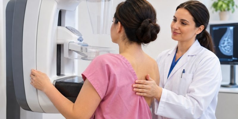
Digital mammography uses low-dose X-rays to create detailed breast images. Unlike older film-based mammograms, digital images can be enlarged and adjusted to help radiologists examine suspicious areas.
This technique can identify abnormalities such as masses, unusual tissue changes,s and tiny calcium deposits known as microcalcifications.
When is it recommended?
Digital mammography is commonly used for routine screening and for investigating symptoms such as breast lumps, nipple changes, or unusual breast discomfort.
If an abnormality appears, doctors may recommend additional mammographic views or ultrasound for further assessment.
2. Digital Breast Tomosynthesis (3D Mammography)
Digital breast tomosynthesis takes multiple X-ray images from different angles and combines them into thin, reconstructed sections of breast tissue.
This allows radiologists to examine individual tissue layers rather than relying entirely on overlapping structures in conventional mammograms.
It can be particularly useful for examining dense breasts, where overlapping tissue may hide small abnormalities or create suspicious-looking areas.
People researching advanced breast imaging may encounter tomosynthesis because it can improve cancer detection and reduce unnecessary callbacks.
However, it still uses X-rays and cannot replace every other diagnostic examination.
3. Breast Ultrasound
Breast ultrasound uses high-frequency sound waves to create images of structures inside the breast. It does not involve ionising radiation and is commonly used alongside mammography.
One important benefit is its ability to help distinguish fluid-filled cysts from solid masses.
For example, if a mammogram identifies a rounded abnormality, ultrasound may help determine whether it contains fluid or solid tissue.
Ultrasound is also useful for investigating palpable lumps and guiding certain breast biopsies.
Although valuable, it does not replace mammography because some abnormalities, particularly tiny calcifications, are better identified using X-rays.
4. Breast Magnetic Resonance Imaging (MRI)
Breast MRI uses magnetic fields and radio waves to produce detailed images of breast tissue. Diagnostic breast MRI commonly involves an injected contrast agent that helps highlight suspicious areas.
Doctors may recommend MRI when conventional imaging provides insufficient information or when someone has a particularly high risk of developing breast cancer.
It can also help assess the extent of disease in selected patients who already have a confirmed diagnosis.
Unlike mammography, MRI does not use ionising radiation.
However, MRI can identify abnormalities that turn out to be non-cancerous, sometimes leading to additional investigations. Its use should therefore depend on individual clinical needs.
5. Contrast-Enhanced Mammography
Contrast-enhanced mammography combines standard mammographic imaging with an iodine-based contrast agent injected into a vein.
The contrast helps radiologists identify areas showing unusual enhancement, which may indicate increased blood supply associated with certain tumours.
This technique can provide additional information when conventional mammograms produce unclear results.
It may also be considered for selected patients who need further assessment but cannot undergo breast MRI.
Because the examination involves iodine-based contrast and X-rays, doctors must consider factors such as previous contrast reactions, kidney function, and pregnancy before recommending it.
6. Computer-Aided Detection (CAD)
Computer-aided detection uses software to analyse mammographic images and flag areas that may require closer examination.
These systems can draw attention to suspicious masses, calcifications, or unusual tissue patterns.
For example, a radiologist may examine a mammogram independently before reviewing areas highlighted by the software.
However, CAD is an additional image-analysis tool rather than a separate scan or a replacement for professional interpretation.
Its accuracy and clinical benefits vary between systems, and flagged areas do not necessarily indicate cancer.
Healthcare facilities such as cleveland clinic abu dhabi describe computer-aided detection among the technologies used in breast imaging assessment.
7. MRI-Guided Breast Biopsy
An MRI-guided breast biopsy is different from the previous techniques because it collects tissue rather than simply producing images.
Doctors use MRI to locate suspicious areas that may not be visible through mammography or ultrasound.
During the procedure, a radiologist uses MRI guidance to position a biopsy needle accurately and remove small tissue samples.
These samples are examined in a laboratory to determine whether cancer cells are present.
MRI-guided biopsy is particularly useful when an abnormality is visible only on MRI and requires tissue confirmation.
How Do Doctors Decide Which Breast Imaging Test Is Necessary?
Choosing an imaging examination depends on several factors. These include the patient’s age, breast density, symptoms, personal risk factors, and previous results.
For example, a newly detected lump may require diagnostic mammography and targeted ultrasound, whereas someone with a high inherited risk of breast cancer may also need MRI.
Doctors generally consider:
- The location and characteristics of a suspicious area.
- Breast density and previous mammogram findings.
- Personal or family history of breast cancer.
- Pregnancy, contrast allergies, or other medical considerations.
- Whether a biopsy is required to confirm the findings.
Patients should also understand their imaging report, particularly the BI-RADS assessment category, which helps explain the level of concern and recommended follow-up.
Conclusion
Advanced breast imaging techniques provide different ways to investigate possible breast cancer, from detailed mammograms and ultrasound examinations to contrast-enhanced scans and MRI-guided biopsies.
The most appropriate investigation depends on individual circumstances rather than using every available technology.
Importantly, an unusual imaging result does not automatically mean cancer. Following recommended appointments, discussing findings with a qualified specialist and completing any necessary biopsy can help establish an accurate diagnosis and determine the next steps.